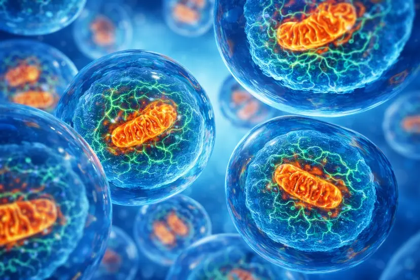
Origins and Evolution of Mitochondria Mitochondria were first described morphologically by Richard Altmann in 1890, who referred to them as “bioblasts,” but their evolutionary origins were clarified much later. In 1967, Lynn Margulis (then Sagan) articulated the endosymbiotic theory, proposing that mitochondria arose from an ancestral α‑proteobacterium engulfed by a proto-eukaryotic cell, explaining the presence […]
Origins and Evolution of Mitochondria
Mitochondria were first described morphologically by Richard Altmann in 1890, who referred to them as “bioblasts,” but their evolutionary origins were clarified much later. In 1967, Lynn Margulis (then Sagan) articulated the endosymbiotic theory, proposing that mitochondria arose from an ancestral α‑proteobacterium engulfed by a proto-eukaryotic cell, explaining the presence of mitochondrial DNA (mtDNA), ribosomes, and bacterial-like inner membranes. Modern imaging and biochemical analyses now show that mitochondria form highly dynamic networks—rather than isolated organelles—within most cell types, continuously undergoing fission and fusion and thereby coordinating energy production, stress responses, and cell fate decisions.
Core Mitochondrial Functions
Mitochondria are often termed the “powerhouses of the cell” because they generate ATP through oxidative phosphorylation, but their functional repertoire is much broader. Key roles include:
- Energy metabolism: Oxidation of carbohydrates, fatty acids, and some amino acids via the tricarboxylic acid (TCA) cycle and β‑oxidation, coupled to ATP production by the electron transport chain (ETC).
- Calcium handling: Buffering and releasing calcium to shape cytosolic Ca²⁺ signals, thereby influencing gene expression, secretion, and cell death pathways.
- Steroidogenesis: Provision of cholesterol transport and early steps in steroid hormone synthesis in specialized tissues (e.g., adrenal cortex, gonads).
- Cell death and signaling: Regulation of apoptosis via cytochrome c release, and participation in innate immune signaling and redox-sensitive pathways.
Mitochondrial Biogenesis and Mitophagy
Mitochrial biogenesis is the process by which cells increase mitochondrial mass and number, requiring coordinated expression of nuclear and mitochondrial genomes. The transcriptional coactivator peroxisome proliferator-activated receptor-γ coactivator-1α (PGC‑1α) is a central regulator that integrates signals from exercise, fasting, cold exposure, and oxidative stress to drive mitochondrial gene expression, mtDNA replication, and assembly of respiratory complexes. PGC‑1α acts through downstream transcription factors including NRF1/NRF2 and mitochondrial transcription factor A (TFAM), which together orchestrate mitochondrial protein synthesis, membrane formation, and respiratory chain assembly. Biogenesis is tightly coupled to mitophagy, the selective autophagic removal of damaged mitochondria, ensuring renewal of a functionally competent mitochondrial pool over time.
Mitochondria and Metabolic Flexibility
Metabolic flexibility—the ability to switch between lipid and carbohydrate oxidation according to substrate availability and energy demand—is largely mediated by mitochondria. During fasting or ketogenic states, mitochondrial β‑oxidation of fatty acids generates acetyl‑CoA for the TCA cycle and, in the liver, ketone bodies that serve as alternative fuels for brain and muscle. Under carbohydrate-rich conditions, pyruvate derived from glycolysis is oxidized via pyruvate dehydrogenase, with malonyl‑CoA and carnitine palmitoyltransferase-1 (CPT1) acting as key regulators of fatty acid entry into mitochondria. Mitochondria also function as energy sensors: falling ATP/AMP ratios activate AMP‑activated protein kinase (AMPK), which promotes catabolic, ATP-generating pathways and inhibits mechanistic target of rapamycin (mTOR), thereby restraining growth and favoring energy conservation. Over longer timescales, sustained energetic demand (e.g., endurance training) can upregulate mitochondrial biogenesis to support improved oxidative capacity and fatigue resistance.
Mitochondria, Redox Balance, and Aging
Redox reactions underlie mitochondrial energy metabolism, with the ETC serving as a major site of reactive oxygen species (ROS) generation. While excessive ROS can damage lipids, proteins, and nucleic acids, low to moderate levels of mitochondrial ROS act as signaling molecules in processes such as insulin signaling and adaptation to exercise. Imbalance between ROS production and antioxidant defenses leads to oxidative stress, which can damage mtDNA—located near the ETC and lacking the full protective chromatin structure and repair capacity of nuclear DNA—thus promoting further ETC dysfunction and ROS production in a self-amplifying cycle. High ROS can also react with nitric oxide to form reactive nitrogen species, contributing to protein nitration and mitochondrial dysfunction; chronic oxidative stress has been linked to impaired PGC‑1α signaling, diminished mitochondrial biogenesis, and accumulation of dysfunctional mitochondria in aging tissues and age-related diseases.
Mitochondrial-Derived Peptides: MOTS-c and SHMOOSE
MOTS-c
MOTS‑c is a 16–amino acid peptide encoded within the 12S rRNA region of mtDNA and translated in the cytosol, representing one of several mitochondrial-derived peptides (MDPs) with endocrine-like functions. In preclinical models, MOTS‑c improves metabolic homeostasis and insulin sensitivity, particularly in skeletal muscle, largely via activation of AMPK, modulation of folate cycle–linked one‑carbon metabolism, and downstream effects on glucose utilization and stress resistance. MOTS‑c has been shown to translocate to the nucleus under metabolic or oxidative stress, where it regulates stress-response genes and promotes antioxidant defenses, including upregulation of nuclear factor erythroid 2–related factor 2 (NRF2) and its target antioxidant enzymes. Experimental data also suggest that MOTS‑c can enhance markers of mitochondrial quality control—such as fusion-related proteins (MFN2, OPA1) and biogenesis-associated factors (TFAM, NRF1, COX subunits)—although these findings are still largely limited to animal and cell culture studies.
SHMOOSE
SHMOOSE is a recently characterized human MDP encoded by a short open reading frame in mtDNA, identified through integrative genomic and proteomic approaches. Genetic analyses have linked a missense variant in the SHMOOSE-encoding region to an approximately 30% increased risk of late-onset Alzheimer’s disease, suggesting a potential role in neurodegeneration. Functional studies indicate that SHMOOSE localizes to the inner mitochondrial membrane and modulates mitochondrial bioenergetics and neuronal energy signaling, including effects on oxygen consumption rates in neuronal cell models. Ongoing work is investigating SHMOOSE levels in cerebrospinal fluid and their associations with age, white matter volume, and Alzheimer’s disease biomarkers such as tau, with the long-term goal of assessing its utility as a biomarker or therapeutic target.
Implications for Aging and Disease
Mitochondrial dysfunction is strongly implicated in metabolic disorders, type 2 diabetes, cancer, and neurodegenerative diseases, in part through disrupted bioenergetics, impaired mitophagy, and chronic redox imbalance. The discovery of MDPs such as MOTS‑c and SHMOOSE underscores that mitochondria are not only metabolic organelles but also signaling hubs that communicate stress and energetic status to the nucleus and other tissues. These peptides may represent promising experimental leads for targeting aging-related pathophysiology, metabolic syndrome, and neurodegeneration, but clinical translation remains at an early stage.
Nutritional and Supplement-Based Modulation of Mitochondrial Function
Several dietary components and nutraceuticals have been investigated for their ability to support mitochondrial function, often converging on PGC‑1α, AMPK, and antioxidant pathways:
- Resveratrol: Activates SIRT1 and AMPK, thereby enhancing PGC‑1α–dependent mitochondrial biogenesis and improving mitochondrial function in preclinical models.
- Coenzyme Q10 (CoQ10): An essential component of the ETC that supports electron transfer and acts as an antioxidant; supplementation may benefit conditions associated with mitochondrial dysfunction and statin use.
- Alpha‑lipoic acid (ALA): A redox-active cofactor that can regenerate other antioxidants and has been reported to improve mitochondrial function and insulin sensitivity in some models.
- L‑carnitine: Facilitates mitochondrial fatty acid transport via the carnitine shuttle and may improve fatty acid oxidation in selected mitochondrial and metabolic disorders.
- Creatine: Buffers cellular energy through the phosphocreatine system and can indirectly support mitochondrial energetics, particularly in high‑demand tissues such as muscle and brain.
- B‑vitamins and magnesium: Act as cofactors for numerous mitochondrial enzymes involved in the TCA cycle, oxidative phosphorylation, and one‑carbon metabolism.
- Berberine and EGCG: Plant-derived compounds that activate AMPK and can promote PGC‑1α–linked mitochondrial biogenesis and antioxidant defenses in experimental systems.
Effects of these agents are context dependent and vary across individuals and disease states, and most mechanistic data derive from cell and animal models rather than large, controlled human trials. Nonetheless, collectively they highlight the central role of mitochondrial signaling and redox biology in shaping metabolic health and aging trajectories.
References
- Friedman JR, Nunnari J. Mitochondrial form and function. Nature. 2014;505(7483):335–343.
- Martin WF, et al. Endosymbiotic theories for eukaryote origin. Mol Biol Cell. 2017;28(10):1285–1297.
- Nunnari J, Suomalainen A. Mitochondria: In sickness and in health. Cell. 2012;148(6):1145–1159.
- Giorgi C, et al. Mitochondrial calcium homeostasis as critical factor in cell physiology and disease. Cell Metab. 2018;28(2):265–281.
- Scarpulla RC. Metabolic control of mitochondrial biogenesis through the PGC‑1 family regulatory network. Biochim Biophys Acta. 2011;1813(7):1269–1278.
- Brand MD. Mitochondrial generation of superoxide and hydrogen peroxide as the source of mitochondrial redox signaling. Free Radic Biol Med. 2016;100:14–31.
- Lee C, Zeng J, Drew BG, et al. The mitochondrial-derived peptide MOTS‑c promotes metabolic homeostasis and reduces obesity and insulin resistance. Cell Metab. 2015;21(3):443–454.
- Kim KH, Son JM, Benayoun BA, Lee C. The mitochondrial-encoded peptide MOTS‑c translocates to the nucleus to regulate nuclear gene expression in response to metabolic stress. Cell Metab. 2018;28(3):516–524.e7.
- Anderson J, et al. A mitochondrial microprotein associated with Alzheimer’s disease risk. Mol Psychiatry. 2023;28:1294–1306.
- Skulachev VP, et al. Mitochondrial-targeted antioxidants: Regulation of mitochondrial functions and longevity. Antioxidants. 2024;13(5):613.
- Hood DA, Memme JM, Oliveira AN, Triolo M. Maintenance of skeletal muscle mitochondria in health, exercise, and aging. Annu Rev Physiol. 2019;81:19–41.